Facilities & Scanners
Scanners:
Kim Vang Hansen, September 2020
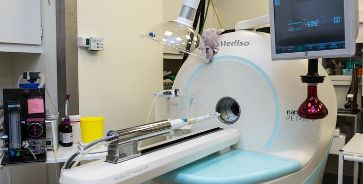
The Mediso nanoScan PET/MRI scanner was delivered from Mediso Medical Imaging Systems in 2013.
The MR part of the system is a self-shielded 1T permanent MR magnet, which does not require cooling. We have two radiofrequency coils, a 35-mm and a 60-mm diameter coil, used for imaging of mice and rats, respectively.
MR imaging can be performed using 2D and 3D sequences such as spin echo and gradient echo. The MR provides good soft tissue contrast required for anatomical/morphological imaging with a spatial resolution down to 0.1 mm.
The PET part consist 12 detector modules, with a total of 38,000 lutetium yttrium oxyorthosilicate (LYSO) crystals of 1.1 x 1.1 x 13 mm3, and magnetically shielded position-sensitive photomultiplier tubes. The PET system has a 9.4 mm (axial) and 12.0 mm (transaxial) coverage. PET data are acquired in list-mode, and several reconstruction methods are available including 3D iterative reconstruction with all corrections including attenuation and scatter in the animal, detector geometry (PSF), depth-of-interaction (DOI), and positron range. The resulting PET images have a high and relatively uniform spatial resolution around 0.7 mm. We currently use the PET/MR-system for small-animal research in brain, kidney and tumour physiology using a variety of PET tracers.
Ole Lajord Munk & Lars Poulsen Tolbod, January 2024
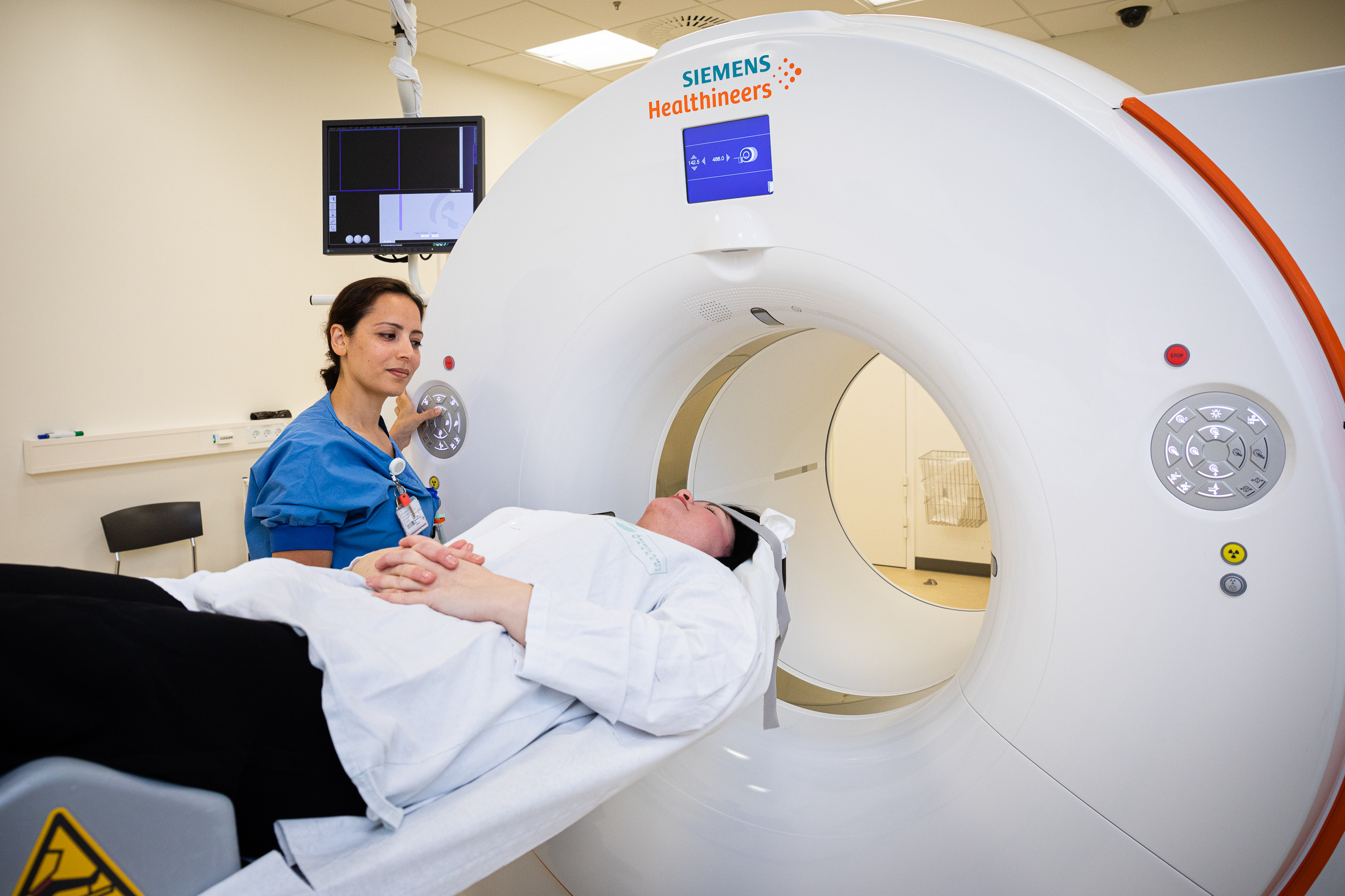
The department’s portfolio of PET-scanners includes a total of six PET/CT scanners: The newest and most advanced is a Siemens Biograph Vision Quadra scanner with a large 106 cm axial field-of-view (installed in 2023). The Quadra scanner has ten times higher sensitivity compared to standard PET/CT scanners and allow simultaneous imaging of all major organs. The scanner is mainly used for imaging of children and young adults as well as research.
The remaining five standard-field-of-view scanners consists of three Siemens Biograph Vision 600 scanners, one GE Discovery MI-5 scanner and one GE Discovery MI Digital-Ready. The Vision and MI-5 scanners have PET detectors based on silicon photomultiplier technology.
The three Vision scanners from Siemens Healthcare are equipped with 128 slice CT and were installed during 2018-2019. These scanners include a PET detector with 26 cm axial field-of-view and software for multi-parametric imaging.
The two scanners from GE Healthcare are equipped with 128 slice CT and were installed in 2019 (MI-5) and 2017 (MI-DR). The Discovery MI-5 include a PET detector with 25 cm axial field-of-view. The MI-DR scanner include a PET detector with 16 cm axial field-of-view and is used for examinations of single organs, such as heart and brain.
The Siemens Biograph Vision 600 scanners + GE Discovery MI-5 are mainly used for oncological whole-body PET examinations and are equipped with software to correct for respiratory motion for optimal tumor detection.
All PET/CT scanners have time-of-flight PET subsystems and can acquire PET-data in list-mode, giving ultimate flexibility to process data for dynamic studies or to correct for movement using respiratory or ECG gating. The software includes a number of processing options as well as reconstruction algorithms like FBP, OSEM, and OSEM with point spread modelling (PSF). In addition, GE MI-DR and GE MI-5 have a Bayesian Penalized Likelihood reconstruction algorithm (Q.Clear).
Søren Baarsgaard Hansen, September 2020
In 2019 a GE Signa hybrid PET/MR scanner was installed in the department. Compared to the combined PET/CT scanners, the MR part of the system offers anatomical imaging with improved soft-tissue contrast as well as imaging of various functional parameters. The system is equipped with extensive accessories and software packages allowing numerous variants of MR imaging including diffusion imaging, DTI, BOLD, proton spectroscopy, quantification of fat fraction, and MR-elastography. The PET subsystem is a modern silicon photomultiplier based detector technology with time-of-flight (TOF) capability and an 25 cm axial field of view. PET data acquisition takes place in list-mode, and data processing includes various types of MR based attenuation correction and a number of reconstruction algorithms like VUE Point HD/FX and Q.Clear.
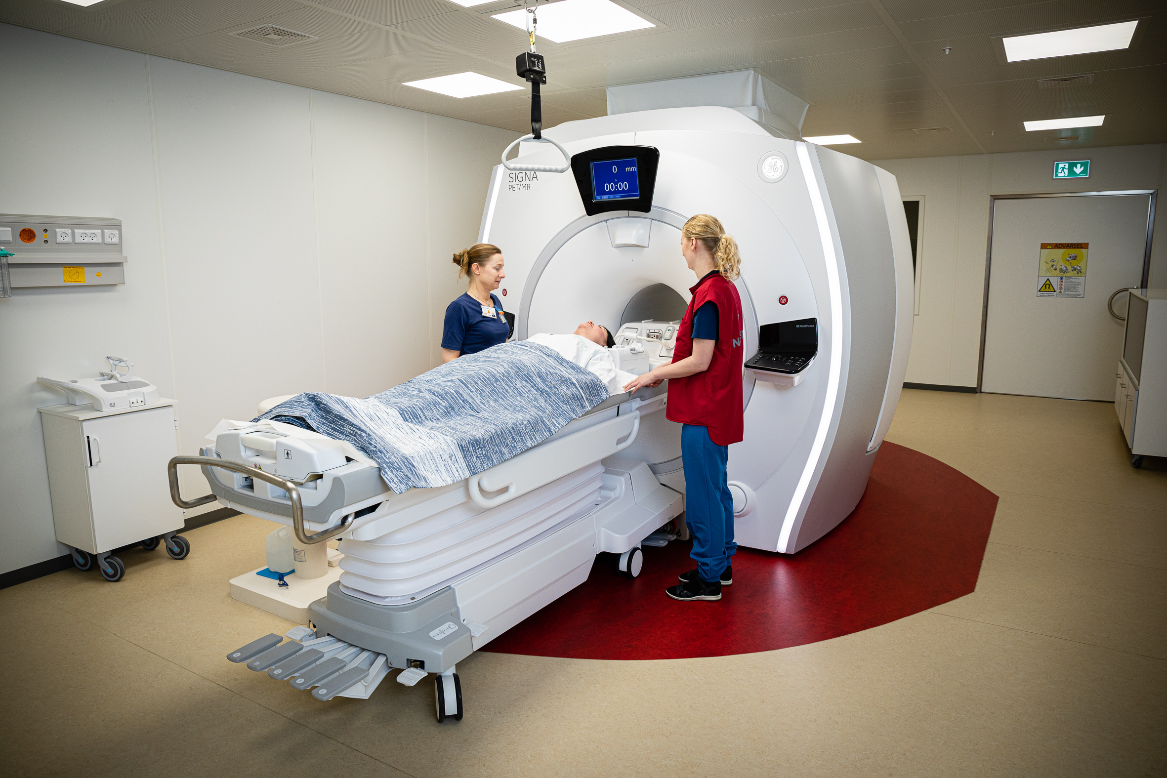
Photo: Tonny Foghmar
Preclinical facilities:
Aage Kristian Olsen Alstrup, September 2022
We have separate surgical facilities for small (mice and rats) and large animals (pigs). For the rodents we have surgical room and a perfusion fixation room. For the large animals we have preparation room, two surgical rooms and a perfusion fixation room for post mortem examinations.
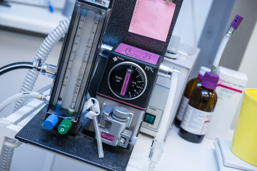
The surgical rooms are used for preparation of animals for imaging studies, but we also have equipment for major surgery procedures, such as implantation of electrodes into the brain or ligation of bile ducts in research of chronic liver diseases. These two surgical rooms can both be used for preparation of animals prior to imaging studies or used for major surgical procedures. All surgical procedures on pigs not involving imaging, are performed at the farm. All procedure rooms at the farm and at our department, have gas outlets for inhalation anaesthesia.
Tracer production:
Additional instrumentation:
Anne M. Landau, April 2016
PET is a tool for in vivo imaging and can be performed in a longitudinal manner where the same subjects can be scanned at various points in time. This facilitates investigation of disease mechanisms and therapeutic approaches over time.
Autoradiography, on the other hand, can be used to visualise and quantify in vitro densities of specific target proteins without competition from endogenous ligands. Autoradiography is performed only at one time-point since it requires postmortem tissue processing. However, it has the advantage of avoiding confounding effects of tracer metabolism, blood flow, passage through the blood brain barrier and plasma protein binding which can complicate PET measurements. Parameters such as binding potential and dissociation constant can be quantified using autoradiography, which can be useful in the characterization of new potential PET ligands.
A wide range of radioligands of specific targets is available for autoradiography. We routinely use [3H], [125I], [11C] and [18F] radioligands in rodent, pig and human brain and peripheral organ fresh frozen tissue sections to quantify receptor and transporter binding and protein aggregation in different disease states. In rodent studies, experiments can also be performed ex vivo, after injection of a radiolabelled tracer into the animal, followed by brain or tissue removal and processing. Studies are currently performed using a BAS-5000 image reader with a resolution of 50 mm and Fujifilm imaging plates. In the near future, it is our intention to replace this system with a new beta imager as a safer, faster and more cost-efficient alternative.
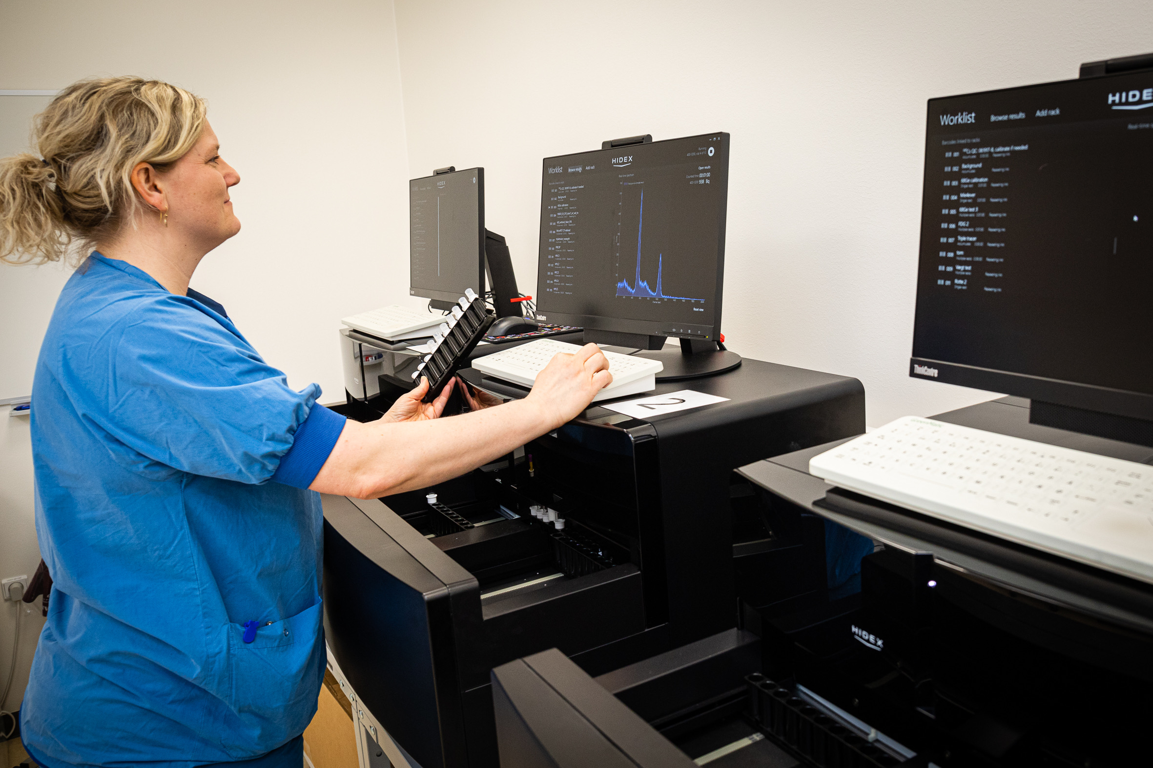
Hidex AMG well counters
Ole Lajord Munk, September 2020
For research purposes, we often supplement PET measurements for quantitative studies with automated or manual blood sampling. For automated sampling, we have four automated blood sampling system. Two Allogg ABSS that use a BGO crystal for efficient detection of radiation from PET tracers in the blood – these are mainly used for blood sampling in humans and pigs. We also have two SwissTrace Twilite Three with LYSO detectors that are MR compatible and can be used with an arterio-venous shunt in small animals to allow measurements without any blood loss. Blood is withdrawn using a one-directional peristaltic pump and allows a dense and continuous (1-second steps) measurement of the time course of the activity concentration in the blood; this is preferred when the tracer has very fast kinetics, e.g. for perfusion measurements. We use dedicated software to provide calibrated data, which is corrected for dispersion and delay caused by the tubes, to obtain true arterial time-activity curves. Alternatively, our experienced technicians can manually withdraw 1-2 mL blood samples by syringes every 5 sec. An advantage of manual blood sampling is that plasma samples can be extracted, which is required for tracers where activity concentrations are different in plasma and blood. Manual blood sampling is also required in metabolite analyses. Radioactivity concentrations of manual blood/plasma samples are measured using our three Hidex AMG well counters that handle multiple racks of 10 test tubes. All PET scanners are cross-calibrated with our blood sampling equipment to allow easy analyses when combining PET data and blood data.
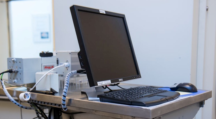
Allogg automated blood sampling system
