Functional CT (DCE-CT) and dual detector CT
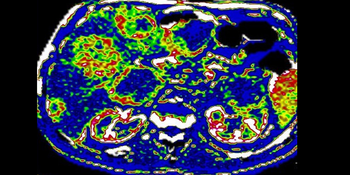
For years, the department has worked with the development of functional imaging. There is no well described protocol in functional CT, and the research group within functional CT uses phantoms and animal models to study the fundamentals of dynamic contrast enhanced CT (DCE-CT) as CT acquisition parameters and contrast media kinetics, to obtain the most optimal protocol in the DCE-CT discipline. This is important when DCE-CT results are compared across institutions.
Functional parameters are important adjuncts to morphologic features in the prognostics and tumour characterisation as well as in follow up of cancer therapy. Both local cancer treatment such as RF-ablation, chemoembolisation and radiation therapy, and systemic cancer treatment such as immunotherapy and targeted therapy are investigated.
Different DCE-CT parameters and figures of image texture analysis are tested and correlated to histopathology and/or end points as time to progression, progression free survival etc.
Spectral CT data seems to have an impact in detecting and characterising lesions. Studies comparing perfusion figures and spectral data figures are investigated.
References
- Mains JR, Donskov F, Pedersen EM, Madsen HHT, Thygesen J, Thorup K, Rasmussen F. Use of patient outcome endpoints to identify the best functional CT imaging parameters in metastatic renal cell carcinoma patients. Br J Radiol. 2018 Feb;91(1082):20160795. doi: 10.1259/bjr.20160795. Epub 2018 Jan 2. PubMed PMID: 29144161; PubMed Central PMCID: PMC5965772.
- Mains JR, Donskov F, Pedersen EM, Madsen HH, Rasmussen F. Dynamic Contrast-Enhanced Computed Tomography-Derived Blood Volume and Blood Flow Correlate With Patient Outcome in Metastatic Renal Cell Carcinoma. Invest Radiol. 2017 Feb;52(2):103-110. doi: 10.1097/RLI.0000000000000315. PubMed PMID: 27513367.
- Mains JR, Donskov F, Pedersen EM, Madsen HH, Rasmussen F. Dynamic contrast-enhanced computed tomography as a potential biomarker in patients with metastatic renal cell carcinoma: preliminary results from the Danish Renal Cancer Group Study-1. Invest Radiol. 2014 Sep;49(9):601-7. doi: 10.1097/RLI.0000000000000058. PubMed PMID: 24691140.
- Andersen IR, Thorup K, Andersen MB, Olesen R, Mortensen FV, Nielsen DT, Rasmussen F. Texture in the monitoring of regorafenib therapy in patients with colorectal liver metastases. Acta Radiol. 2019 Sep;60(9):1084-1093. doi: 10.1177/0284185118817940. Epub 2019 Jan 6. PubMed PMID: 30612433.
- Andersen IR, Olesen R, Boysen AK, Jensen LH, Mortensen FV, Nielsen DT, Rasmussen F. Dynamic contrast-enhanced computed tomography as a potential biomarker in patients with metastatic colorectal cancer treated with regorafenib. Acta Radiol. 2019 Jul;60(7):836-845. doi: 10.1177/0284185118806652. Epub 2018 Oct 22. PubMed PMID: 30348001.
- Andersen IR, Thorup K, Jepsen BN, Mortensen FV, Nielsen DT, Rasmussen F. Dynamic contrast-enhanced computed tomography in the treatment evaluation of patients with colorectal liver metastases treated with ablation: a feasibility study. Acta Radiol. 2019 Aug;60(8):936-945. doi: 10.1177/0284185118806661. Epub 2018 Oct 18. PubMed PMID: 30335477
- Harders SW, Madsen HH, Nellemann HM, Rasmussen TR, Thygesen J, Hager H, Andersen NT, Rasmussen F. Dynamic contrast-enhanced CT in suspected lung cancer: quantitative results. Br J Radiol. 2013 Nov;86(1031):20130257. doi: 10.1259/bjr.20130257. Epub 2013 Sep 12. PubMed PMID: 24029629; PubMed Central PMCID: PMC3830431.
- Harders SW, Madsen HH, Nellemann HM, Rasmussen TR, Thygesen J, Hager H, Andersen NT, Rasmussen F. Can visual assessment of blood flow patterns by dynamic contrast-enhanced computed tomography distinguish between malignant and benign lung tumors? Acta Radiol Open. 2017 May 30;6(5):2058460117710053. doi: 10.1177/2058460117710053. eCollection 2017 May. PubMed PMID: 28607762; PubMed Central PMCID: PMC5453405.
- Andersen MB, Harders SW, Ganeshan B, Thygesen J, Torp Madsen HH, Rasmussen F. CT texture analysis can help differentiate between malignant and benign lymph nodes in the mediastinum in patients suspected for lung cancer. Acta Radiol. 2016 Jun;57(6):669-76. doi: 10.1177/0284185115598808. Epub 2015 Aug 12. PubMed PMID: 26271125.
- Andersen MB, Ganeshan B., Harders SW,Thygesen J, Madsen HT, Rasmussen F. CT texture analysis of pulmonary lesions in patients suspected for lung cancer.Cancer imaging : 2014, the official publication of the International Cancer Imaging Society 14(Suppl 1):S6-S6.DOI: 10.1186/1470-7330-14-S1-S6
- Driljevic-Nielsen A, Rasmussen F, Mains JR, Donskov F. High baseline blood volume is an independent favorable prognostic factor for overall and progression-free survival in patients with metastatic renal cell carcinoma. October 2019, Annals of Oncology 30(Supplement_5); DOI: 10.1093/annonc/mdz249.075.
- Andersen, M.B., Ebbesen, D., Thygesen, J. et al. Impact of spectral body imaging in patients suspected for occult cancer: a prospective study of 503 patients. Eur Radiol 30, 5539–5550 (2020). https://doi.org/10.1007/s00330-020-06878-7
- Drljevic-Nielsen A, Rasmussen F, Mains JR, Thorup K, Donskov F. Baseline blood volume identified by dynamic contrast-enhanced computed tomography as a new independent prognostic factor in metastatic renal cell carcinoma. Transl Oncol. 2020 Oct;13(10):100829. doi: 10.1016/j.tranon.2020.100829. Epub 2020 Jul 9. PMID: 32653813; PMCID: PMC7350156.
Contacts
Finn Rasmussen, MD, DMSc, Associate Professor, Consultant Radiologist: finnrasm@rm.dk
Jesper Thygesen, MSc, PhD student: JESTHY@rm.dk
Stefan Harders, MD, PhD: swharders@gmail.com
Jill Mains, MD, PhD: jillmain@rm.dk
Iben Rahbek Andersen, MD, PhD: Iben.Rahbek.Andersen@rm.dk
Michael Brun Andersen, MD, PhD student: Michael.brun.andersen@regionh.dk
Aska Drijevic-Nielsen, MD, PhD student: askadrl@rm.dk