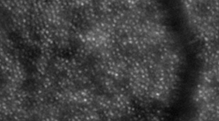
Adaptive optics image of retinal photoreceptors in a healthy person. A retinal arteriole is seen to pass vertically in the right part of the image.
The clinical research unit has special equipment for studying retinal vascular function. The Dynamic Vessel Analyzer allows real time recording of diameter changes of retinal arterioles in vivo during intervention with vasoactive drugs. The reaction of retinal arterioles is studied during an increase in the arterial blood pressure induced by isometric exercise and during stimulation with flickering light which allows quantification of the autoregulation of retinal blood flow. The oxymeter is a modified fundus camera that records images of the ocular fundus at two different wavelengths used to calculate oxygenation of retinal arterioles and venules. Adaptive Optics imaging is a high resolution imaging modality that corrects for imperfections in the imaging through the optics of the eye and thereby allows the resolution of the consequences of vascular disease in the retina in detail.
Reference:
Tilma KK, Bek T. Topical treatment for 1 week with latanoprost but not diclofenac reduces the diameter of dilated retinal arterioles in patientes with type 1 diabetes mellitus and mild retinopathy. Acta Ophthalmol 2011, Jul 5. [Epub ahead of print]
Bek T. Regional morphology and pathophysiology of retinal vascular disease. Prog Ret Eye Res 2013;36:247-59
Contact:
Professor Toke Bek
Phone: +45 7846 3223
E-mail: toke.bek@mail.tele.dk