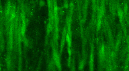
Confocal scanning image of calcium activation in retinal vascular smooth muscle cells during adenosine induced relaxation.
A number of in vitro techniques have been developed for the study of tone regulation in retinal arterioles. The experiments are performed on porcine tissue collected on a daily basis from a nearby slaughterhouse. Techniques have been set up for studying diameter changes in cannulated isolated arterioles using isobaric technique and for studying tone changes in retinal and ciliary arterioles with and without preserved perivascular retinal tissue using isometric technique. Recently, techniques have been developed for perfusion of a whole retinal segment in order to study differences in tone regulation between retinal arterioles with different caliber and disturbances in retinal microcirculation. Additionally, confocal microscopy is used to study the cellular basis for tone regulation in retinal arterioles.
Reference:
Misfeldt MW, Aalkjaer C, Simonsen U, Bek T. Novel cellular bouton structure activated by ATP in the vascular wall of porcine retinal arterioles. Invest Ophthalmol Vis Sci 2010;51/12:6681-7
Kringelholt S, Holmgaard K, Bek T. Relaxation of porcine retinal arterioles during acute hypoxia in vitro depends on prostaglandin and NO synthesis in the perivascular retina. Curr Eye Res 2013;38/9:965-71
Contact:
Professor Toke Bek
Phone: +45 7846 3223
E-mail: toke.bek@mail.tele.dk