
Magnetoencephalography (MEG) in Denmark takes place at the Department of Clinical Neurophysiology at Aarhus University Hospital.
The MEG records from 306 places on the head.
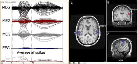
Studies have shown that magnetoencephalography (MEG) is a sensitive technique for localisation of epileptic discharges. MEG records the changes in spontaneous magnetic fields generated by the activity of the neural networks in the brain. The recorded signal arises mainly from the sulci in the brain. In contrast, the electroencephalographic (EEG) signal is mainly generated from the gyri. MEG and EEG used in combination can lead to a better hypothesis about the presumed epileptic focus. It is still debated to which extent MEG yields new clinical information (De Tiege et al. 2012).
Some patients with epilepsy sense the seizure coming before it is detected on the EEG, indicating that physiological changes may happen before the seizure. Physiological changes in Heart Rate Variability (HRV) may be associated with seizures (Jeppesen et al. 2010).
Our research will assess the clinical usefulness of MEG in the diagnostic process of patients in pre-surgical evaluation with therapy resistant focal epilepsy and of patients suspected of epilepsy, but with no EEG proof. Also detection of epileptic seizures will be assessed with HRV to make a portable seizure-alarm. Furthermore, Psychogenic Non Epileptic Seizure will be compared to epileptic seizure using the above methods to distinguish between the two methods for clinical diagnostic purposes.
MEG will be considered for patients with therapy resistant focal epilepsy undergoing pre-surgical evaluation and patients with suspected epilepsy, and patients who have had three non-informative EEGs. Patients will be recruited from the Department of Neurology at Rigshospitalet, The Danish Epilepsy Centre in Dianalund and the Department of Neurology at Aarhus University Hospital.
Simultaneously, MEG and EEG are recorded followed by magnetic resonance imaging (MRI). The epileptic focus found by MEG is superimposed on the MRI. HRV measurements take place continuously when patients are admitted in the video-EEG monitoring unit.
Contact
Sándor Beniczky, MD, PhD, Professor sbz@filadelfia.dk
Lene Duez, MD, leneduez@rm.dk
Jesper Jeppesen, MSc, PhD jespjepp@rm.dk
Peter Orm Hansen, MD, PhD, Head Consultant peter.orm.hansen@aarhus.rm.dk
Anders Fuglsang-Frederiksen, Professor, MD, DMSc andefugl@rm.dk
Mustafa Aykut Kural, Research Assistant, muskur@rm.dk