Scanners
Kim Vang Hansen, March 2025
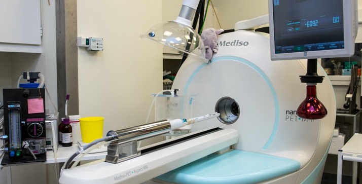
The Mediso nanoScan PET/MRI scanner was delivered from Mediso Medical Imaging Systems in 2013.
The MR part of the system is a self-shielded 1T permanent MR magnet, which does not require cooling. We have two radiofrequency coils, a 35-mm and a 60-mm diameter coil, used for imaging of mice and rats, respectively.
MR imaging can be performed using 2D and 3D sequences such as spin echo and gradient echo. The MR provides good soft tissue contrast required for anatomical/morphological imaging with a spatial resolution down to 0.1 mm.
The PET part consist 12 detector modules, with a total of 38,000 lutetium yttrium oxyorthosilicate (LYSO) crystals of 1.1 x 1.1 x 13 mm3, and magnetically shielded position-sensitive photomultiplier tubes. The PET system has a 9.4 mm (axial) and 12.0 mm (transaxial) coverage. PET data are acquired in list-mode, and several reconstruction methods are available including 3D iterative reconstruction with all corrections including attenuation and scatter in the animal, detector geometry (PSF), depth-of-interaction (DOI), and positron range. The resulting PET images have a high and relatively uniform spatial resolution around 0.7 mm. We currently use the PET/MR-system for small-animal research in brain, kidney and tumour physiology using a variety of PET tracers.
Ole Lajord Munk & Lars Poulsen Tolbod, January 2024
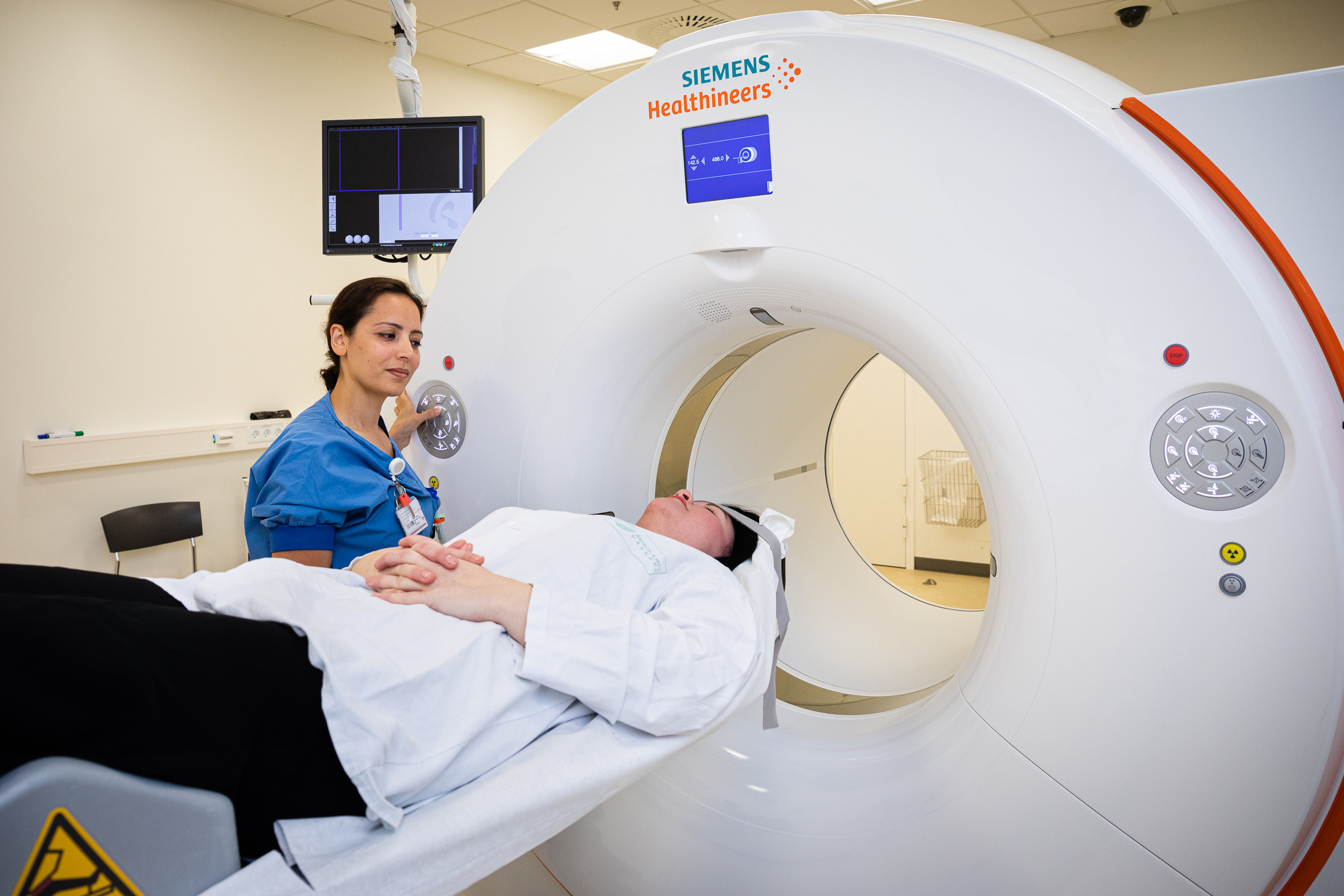
The department’s portfolio of PET-scanners includes a total of six PET/CT scanners: The newest and most advanced is a Siemens Biograph Vision Quadra scanner with a large 106 cm axial field-of-view (installed in 2023). The Quadra scanner has ten times higher sensitivity compared to standard PET/CT scanners and allow simultaneous imaging of all major organs. The scanner is mainly used for imaging of children and young adults as well as research.
The remaining five standard-field-of-view scanners consists of three Siemens Biograph Vision 600 scanners, one GE Discovery MI-5 scanner and one GE Discovery MI Digital-Ready. The Vision and MI-5 scanners have PET detectors based on silicon photomultiplier technology.
The three Vision scanners from Siemens Healthcare are equipped with 128 slice CT and were installed during 2018-2019. These scanners include a PET detector with 26 cm axial field-of-view and software for multi-parametric imaging.
The two scanners from GE Healthcare are equipped with 128 slice CT and were installed in 2019 (MI-5) and 2017 (MI-DR). The Discovery MI-5 include a PET detector with 25 cm axial field-of-view. The MI-DR scanner include a PET detector with 16 cm axial field-of-view and is used for examinations of single organs, such as heart and brain.
The Siemens Biograph Vision 600 scanners + GE Discovery MI-5 are mainly used for oncological whole-body PET examinations and are equipped with software to correct for respiratory motion for optimal tumor detection.
All PET/CT scanners have time-of-flight PET subsystems and can acquire PET-data in list-mode, giving ultimate flexibility to process data for dynamic studies or to correct for movement using respiratory or ECG gating. The software includes a number of processing options as well as reconstruction algorithms like FBP, OSEM, and OSEM with point spread modelling (PSF). In addition, GE MI-DR and GE MI-5 have a Bayesian Penalized Likelihood reconstruction algorithm (Q.Clear).
Søren Baarsgaard Hansen, March 2025
In 2019 a GE Signa hybrid PET/MR scanner was installed in the department. Compared to the combined PET/CT scanners, the MR part of the system offers anatomical imaging with improved soft-tissue contrast as well as imaging of various functional parameters. The system is equipped with extensive accessories and software packages allowing numerous variants of MR imaging including diffusion imaging, DTI, BOLD, proton spectroscopy, quantification of fat fraction, and MR-elastography. The PET subsystem is a modern silicon photomultiplier based detector technology with time-of-flight (TOF) capability and an 25 cm axial field of view. PET data acquisition takes place in list-mode, and data processing includes various types of MR based attenuation correction and a number of reconstruction algorithms like VUE Point HD/FX and Q.Clear.
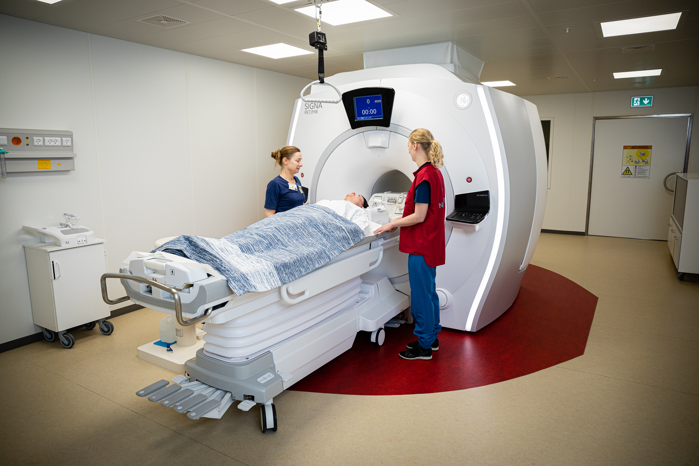
Photo: Tonny Foghmar
Peter Frøhlich Staanum, march 2025
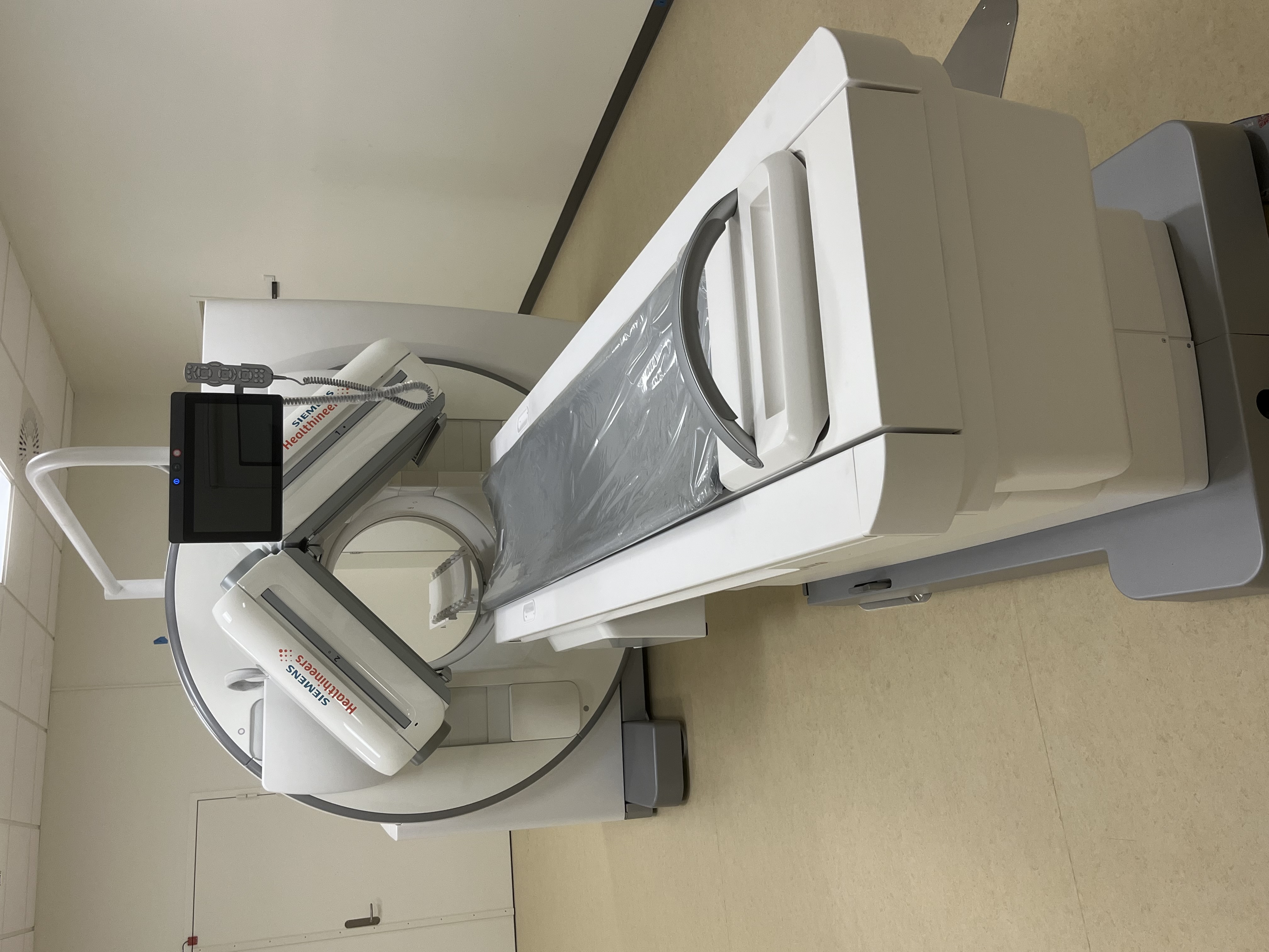
The department operates four SPECT-CT scanners: Two Siemens Symbia T16, one Siemens Symbia Intevo Bold and one Siemens pro.specta X7.
All scanners have low-energy and medium-energy collimators available and three of them also high-energy collimators for imaging of all common diagnostic isotopes (e.g. Tc-99m, In-111, I-123, Se-75) as well as post-radionuclide therapy imaging (I-131, Y-90, Lu-177). The Siemens pro.specta X7 is equipped with a 32-slice CT scanner and contrast-enhanced diagnostic Ct scans are performed here, while the remaining CT-scanners are 16-slice.
SPECT reconstructions can be performed by the proprietary reconstruction software Flash3D or Flash 3D+ (pro.specta), or using the Hybrid Recon application of the vendor-independent Hermes platform. In all cases a number of standard options are available, e.g. FBP and OSEM reconstructions with attenuation correction, scatter correction as well as point-spread-function correction.
Preclinical facilities
Aage Kristian Olsen Alstrup, September 2022
We have separate surgical facilities for small (mice and rats) and large animals (pigs). For the rodents we have surgical room and a perfusion fixation room. For the large animals we have preparation room, two surgical rooms and a perfusion fixation room for post mortem examinations.
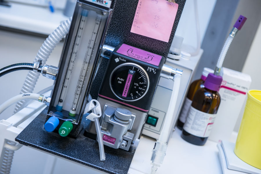
The surgical rooms are used for preparation of animals for imaging studies, but we also have equipment for major surgery procedures, such as implantation of electrodes into the brain or ligation of bile ducts in research of chronic liver diseases. These two surgical rooms can both be used for preparation of animals prior to imaging studies or used for major surgical procedures. All surgical procedures on pigs not involving imaging, are performed at the farm. All procedure rooms at the farm and at our department, have gas outlets for inhalation anaesthesia.
Tracer production
Dirk Bender, March 2025
Traditional nuclear medicine and PET both use diagnostic radioactive pharmaceuticals, called tracers. PET tracers are labelled with radioactive atoms which decay under positron (antiparticles of electron) emission.
Isotopes (used atoms) have a typical short half-life. 2 minutes for 15O and almost 2 hours for 18F. This short half-life means that tracers must be produced on site.
Tracers labelled with short-lived isotopes are singly produced - one tracer per patient. Tracers with a longer half-life are produced for groups of patients.
The goal of the radiochemistry/radiopharmacy unit, is to produce the necessary tracers for clinical and pre-clinical procedures.
Once the radioactive isotopes are produced, using a cyclotron, they are bound to larger molecules, which eventually become the radioactive tracers.
Production laboratories are all classified as a clean-rooms.
After production, and before use, all products are tested in the quality control laboratory, to ensure the specified quality standards. If the tracers meet the quality control requirements, they are released and prepared for distribution.
The radiopharmacy units meets the requirements of the Danish Medicines Agency regarding aseptic production of radioactive pharmaceuticals, and follows the principles of the Good Manufacturing Practice (GMP).
Each year, the PET Centre’s radiopharmacy unit produces approximately 16,000 doses of more than 30 different tracers which are used for human body scans and each year it is aimed to develop 2 – 3 new tracers.
Production starts at 4 a.m. and more than 30 people, including 9 chemists, are involved in the manufacturing process.
The radioactive isotopes are produced with the help of 3 cyclotrons (GE PETtrace and IBA 18/18) all year round.
Ga-68 generators are also used as well.
Production takes place in 4 radiochemistry production laboratories. The laboratories have 16 hot cells which contain 7 synthesis units for F-18 chemistry, 4 units for C-11 chemistry, 1 unit for Ga-68 chemistry and separate modules for O-15
chemistry.
Quality control is maintained by 5 Radio-HPLCs, 1 Radio-LCMS, 1 Radio TLC and 2 GCs.
The tracers are used in clinical and research studies. Further 65 tracers can be produced at short notice for pre-clinical scans.
The radiopharmacy unit produces around 7,500 doses each year in 2 radiochemistry labs with the help of Tc-99 generators and pre-fabricated kits. In addition to the nuclear medicine standard products, the following are also produced: Y-90 and Lu-177 Dotatoc for therapy, C-14 Palmitat and H-3 Glucose. Y-90 Sirtec, Lu-177 Lutathera, Lu-177 PSMA I&T and XOFIGO are used for therapy purposes as well.
Distribution of tracers for clinical use
In 2002, the Danish Medicines Agency granted a marketing authorization of the most widely-used tracer, fluorine-18 FDG. FDG is delivered to PET centres at Aalborg University Hospital, Odense University Hospital, Aarhus University Hospital, Skejby, Herning Regional Hospital and Vejle Regional Hospital.
The tracers NaF, FDOPA, Fluorocholine, FPSMA-1007 and F-18 PE2I are distributed to other hospitals.
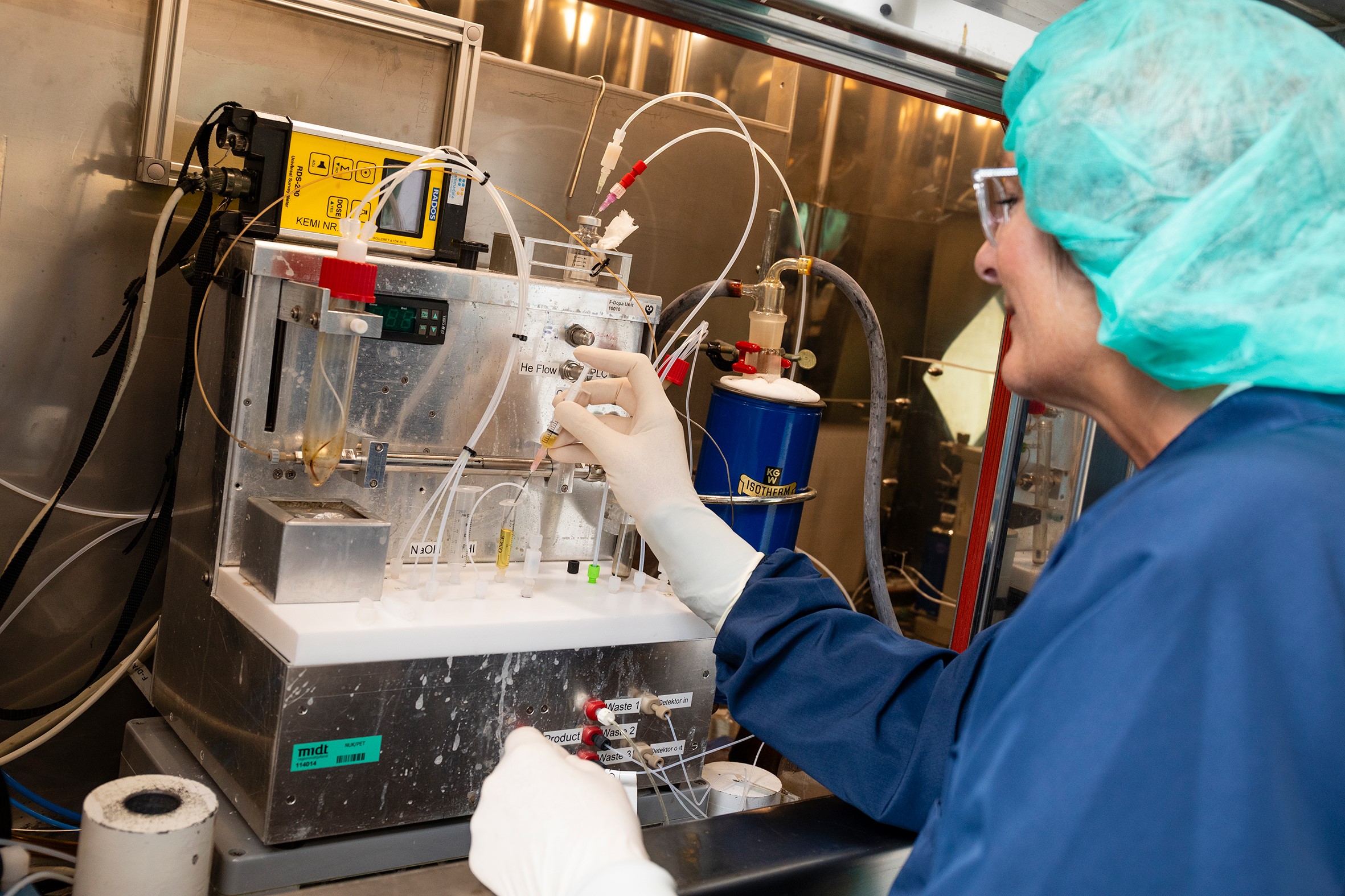
Photo: Tonny Foghmar
Christian Flø, March 2025
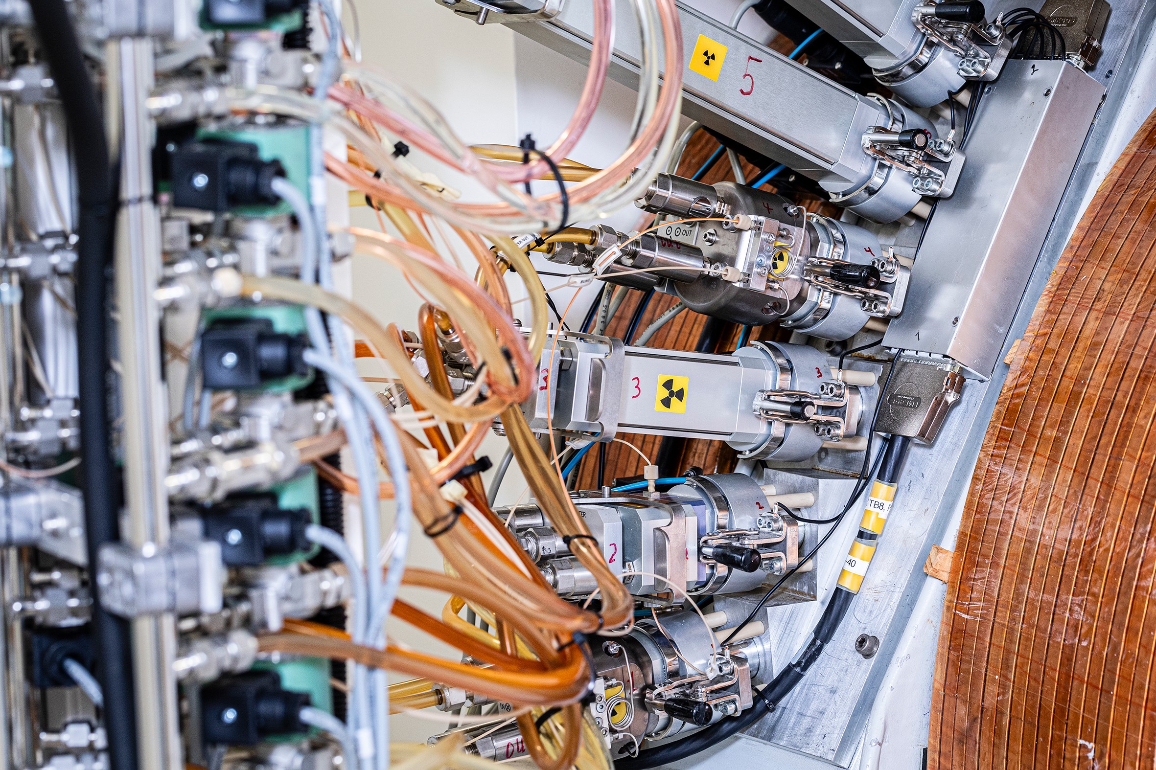
For production of short-lived PET radionuclides, the department operates three medium-sized cyclotrons, two PETtrace systems from GE Healthcare and a Cyclone 18/18 from IBA Radiopharma Solutions. The first PETtrace cyclotron was installed just before the inauguration of the PET-center in Aarhus in 1993, and a second cyclotron, the Cyclone 18/18, was installed in 2009. In 2019 both cyclotrons were moved to the unified Aarhus University Hospital in Skejby and reinstalled after substantial upgraded of various subsystems and targets. In addition, a new PETtrace 880 was installed at the new location.
The PETtrace machines are equipped with six targets, and the acceleration system can deliver beams of protons and deuterons at energies 16,5 and 8,4 MeV respectively. The Cyclone 18/18 cyclotron is equipped with eight targets and is capable of delivering protons at an energy of 18 MeV. The cyclotrons are used routinely for production of the radionuclides 11C, 13N, 15O og 18F, of which several can be delivered as various precursors via online chemical processing modules. All cyclotrons are installed in concrete bunkers including separate maze entrances.
Additional instrumentation:
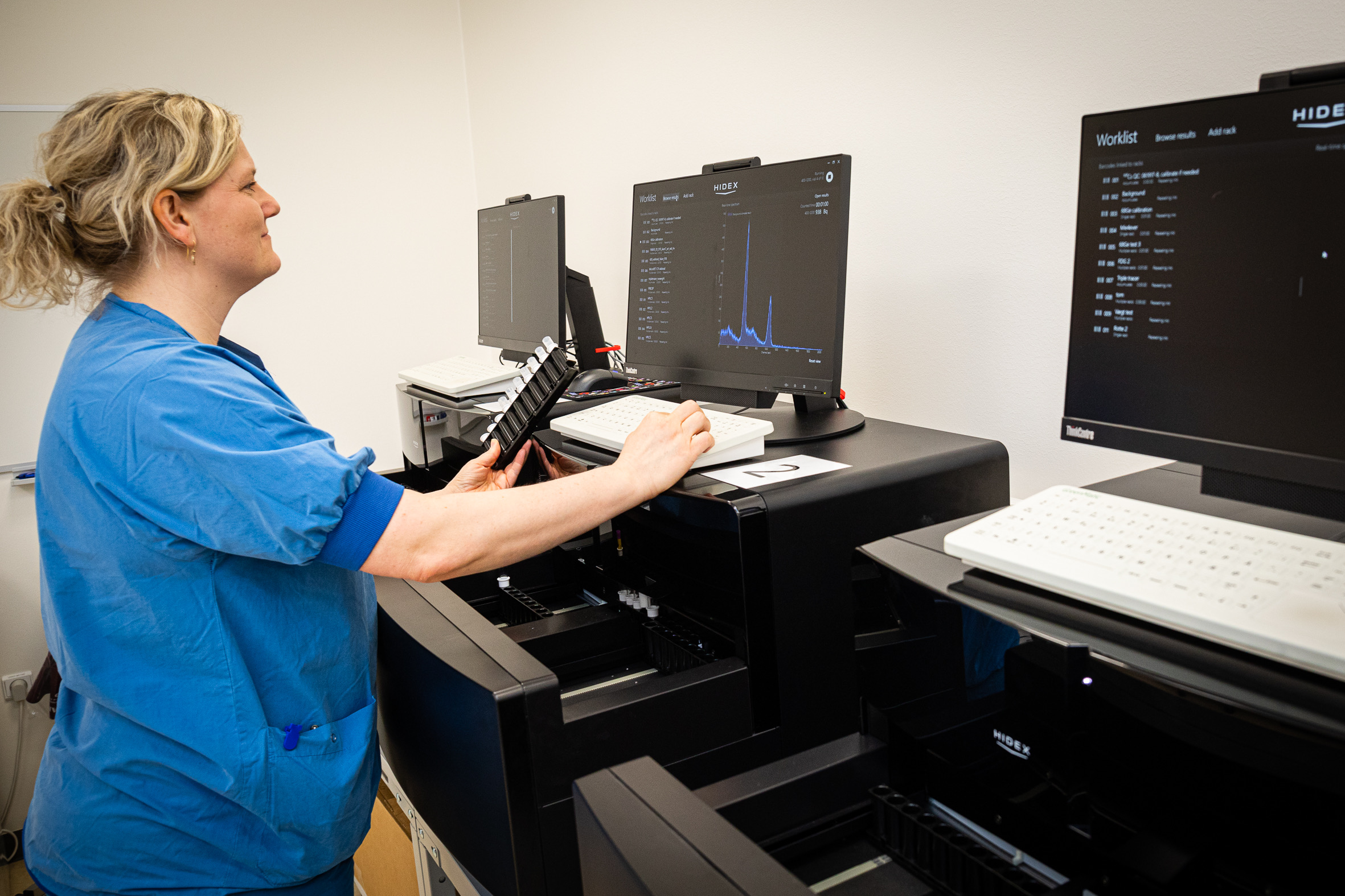
Hidex AMG well counters
Ole Lajord Munk, September 2020
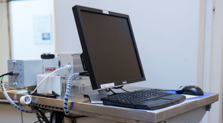
For research purposes, we often supplement PET measurements for quantitative studies with automated or manual blood sampling. For automated sampling, we have four automated blood sampling system. Two Allogg ABSS that use a BGO crystal for efficient detection of radiation from PET tracers in the blood – these are mainly used for blood sampling in humans and pigs. We also have two SwissTrace Twilite Three with LYSO detectors that are MR compatible and can be used with an arterio-venous shunt in small animals to allow measurements without any blood loss. Blood is withdrawn using a one-directional peristaltic pump and allows a dense and continuous (1-second steps) measurement of the time course of the activity concentration in the blood; this is preferred when the tracer has very fast kinetics, e.g. for perfusion measurements. We use dedicated software to provide calibrated data, which is corrected for dispersion and delay caused by the tubes, to obtain true arterial time-activity curves. Alternatively, our experienced technicians can manually withdraw 1-2 mL blood samples by syringes every 5 sec. An advantage of manual blood sampling is that plasma samples can be extracted, which is required for tracers where activity concentrations are different in plasma and blood. Manual blood sampling is also required in metabolite analyses. Radioactivity concentrations of manual blood/plasma samples are measured using our three Hidex AMG well counters that handle multiple racks of 10 test tubes. All PET scanners are cross-calibrated with our blood sampling equipment to allow easy analyses when combining PET data and blood data.
Allogg automated blood sampling system