
The Department of Histopathology is the main provider of diagnostic histopathological and cytopathological services in the Central Denmark Region and acts as a regional and national reference centre. We have substantial teaching and research duties.
We have a large on-site laboratory, housing state-of-the-art automated equipment for use in a wide range of diagnostic methods applied to cells and tissue from patient samples.
Annually, we receive more than 60,000 requests, examine more than 500,000 glass slides by microscopy and perform some 3,500 molecular analyses. A large part of our diagnostic work involves the investigation and treatment of cancer. Additional important areas of specialization include transplantation pathology and the diagnosis of non-neoplastic diseases of the major organs.
The department employs 23 consultant/staff specialist pathologists, has between 11 and 17 residents in training, 80 biomedical laboratory technicians, 4 molecular biologists, 6 secretaries, 1 administrative employee and 6 mortuary staff members.
Routine daily tasks involve:
-
cytology
-
diagnostic molecular pathology
-
histology
-
autopsies
Cytology:
Diagnoses are made based on the microscopic examination of clinical samples consisting mainly of cells. These cells are obtained either from collected body fluids (e.g. urine or pleural fluid) or by the use of fine-needle aspiration or brushing techniques.
Cytology is widely used as a cancer-screening tool. In addition, being a minimally invasive method, it is well-suited for obtaining material from hard-to-reach lesions or from patients who do not tolerate more invasive methods. We perform endo-bronchial ultrasound examinations (EBUS with ROSE) to secure adequate cytological microscopy and molecular analyses.
Diagnostic molecular pathology:
We make use of a wide range of state-of-the-art molecular methods. These allow for a more precise classification of various cancers and enable us to identify specific genetic and epigenetic changes that underlie tumour growth. By combining more precise histological and cytological tumour classification with the molecular analysis of targeted gene panels, it is possible to predict the treatment strategies that are best suited to the individual patient.
Histology:
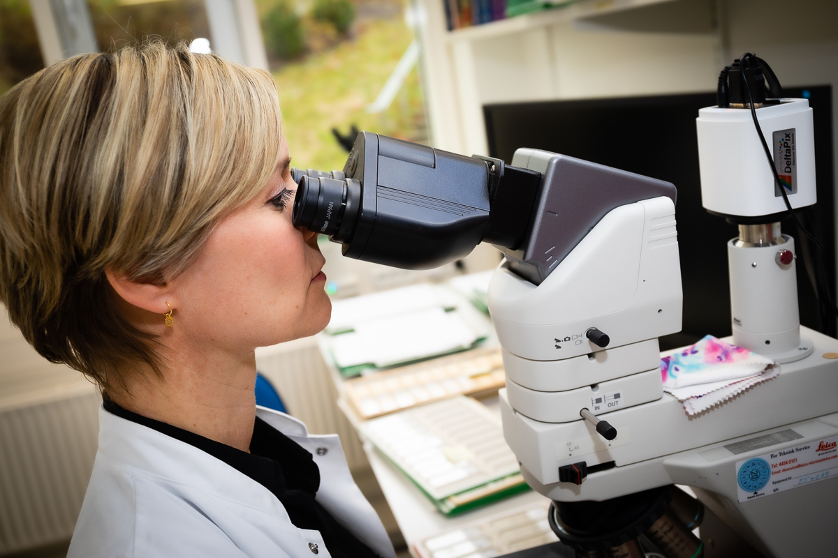
Histopathological diagnosis is based on the microscopic examination of tissue samples, either in the form of small tissue biopsies, or as tissue sections selected macroscopically from large organ specimens removed at surgery. Tissue samples are fixed in formalin and prepared for embedding in paraffin, then cut in thin sections and placed on microscope slides. The slides are then stained to make the cells visible under magnification. At present, the pathologists examine these tissue slides using a traditional microscope; in the future slides will often be digitally scanned and examined on an electronic screen. Digitalization of the slides opens up possibilities for additional analyses based on machine learning/artificial intelligence.
Often, it is necessary to carry out special staining methods. The most important of these staining techniques is immunohistochemistry in which panels of marked antibodies against specific antigens are used to describe tumour or gene specific expression. Knowledge of the marker expression pattern shown by pathological cells or tissues enables more precise diagnosis and may provide prognostic or predictive information of therapeutic value.
If there is need for more rapid diagnosis, it is possible to perform intraoperative frozen section analysis. This may be carried out while the patient is still anaesthetized during an operation, for example to determine whether a cancer has been removed completely, allowing the surgeon the possibility to remove more tissue if necessary. In transplantation pathology, same-day diagnosis is often required, in which case rapid tissue preparation methods can be used, and the pathologist can give a rush diagnosis by telephone.
Autopsies
We perform around 100 hospital autopsies (adults) and around 80 foetus autopsies yearly. The post-mortem room is close to the chapel.
The mortuary staff is responsible for arranging viewing of the deceased to relatives.
We handle more than 1,600 deceased individuals every year.