
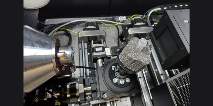
Figure: Sneak peek into the spectral microCT scanner, with the x-ray source on the left, the scanning object (mouse tumour in a cuvette) in the middle, and the Medipix3 detector on the right.
When a patient is diagnosed with cancer, one of the first questions to ask is if the disease is local, and if it can be treated with local curative therapy, such as surgery or radiotherapy. Medical imaging with CT, MR and PET scanners is acquired to answer this question. We are good at localising a tumour on imaging, but it is much more challenging to exactly determine the extent of a tumour. Where the extent of the tumour is the starting point for designing the local treatment plan, any error in segmenting the tumour extent on imaging will directly affect the treatment outcome. In our research, we aim to improve the accuracy with which we can segment tumours on medical imaging. For this we have two main research tracks.
The first is interactive deep-learning based image segmentation. Where deep-learning technology provides us with the best tumour segmentations to date, the quality is not yet at a state that we can fully automate this process. In our research, we update the deep-learning models while the treating physician is assessing the images, providing an accurate tumour segmentation with minimal interaction. This work is aimed at providing clinically useful and accepted deep-learning tools for segmentation by putting the doctors in control.
Our second research track is aimed at better understanding the medical imaging, to know what is tumour, and what not. For this, we have developed a spectral microCT scanner, which is based on a Medipix3 detector. The novelty of this scanner is that the we can obtain the spectrum of the x-rays that pass through a tumour. This spectral information is expected to be different between tumour and surrounding normal tissues, providing more imaging contrast. The goal is to obtain high-resolution (50-100 µm voxels) 3D images of tumours that are removed during surgery, and compare them to pre-operative imaging.
Our research is interdisciplinary, combining physics, computer science, engineering and many different medical specialities, including radiology, radiotherapy, pathology and surgery.
People
 Associate professor of medical physics
Jasper Albertus Nijkamp
jaspernijkamp@clin.au.dk
Further information.
PhD Student
Morten Sahlertz
MORTSA@rm.dk
Collaborating researchers
 Professor of medical physics
Stine Korreman
stkorr@rm.dk
Further information.
Associate professor, the Dep. of Pathology
Trine Tramm
tramm@clin.au.dk
PURE, Aarhus University
Professor, Dep. of Plastic and Breast surgery
Peer Christiansen
peer.christiansen@clin.au.dk
PURE, Aarhus University
Professor, Dep. of Oncology
Jesper Grau Eriksen
jesper@oncology.au.dk
Pure, Aarhus University
PhD student, Dep. of Radiology
Irina Palimaru Manhoobi
irina.palimaru@clin.au.dk
PURE, Aarhus University
Associate professor, Dep. of Otorhinolaryngology
Thomas Kjærgaard
thokjaer@rm.dk
PURE, Aarhus University